

The Key Takeaways:
Typical adult cervical range of motion runs about 45-60 degrees of flexion, 50-70 degrees of extension, 65-75 degrees of rotation to each side, and 35-45 degrees of lateral flexion to each side. The atlanto-axial joint alone supplies roughly half of that rotation, you can get a rough read on your own mobility with a mirror, a phone app, or a simple functional test, and a lost range of motion usually traces back to posture, sleep position, repetitive movement, age, or injury rather than one dramatic cause.
Turning your head to check a blind spot while driving, nodding along in a conversation, or looking up at a top shelf all draw on the same pool of cervical mobility. When that pool shrinks, even small, ordinary motions start to feel effortful. A restricted range of motion can also feed into headaches, muscle imbalances, and extra strain on the shoulders and upper back, which is often how people notice that something has changed before they notice why.
How does the cervical spine allow this much movement?
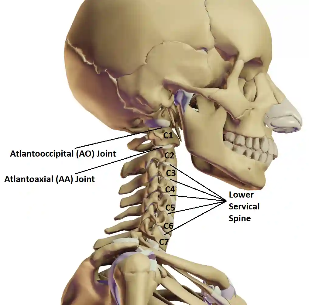
Seven vertebrae, C1 through C7, stack under the skull to support its weight while keeping it mobile. Three structures share that work: the vertebrae themselves provide the structural support and the joint surfaces that let one bone articulate against the next; the intervertebral discs between them cushion the stack and allow smooth gliding motion; and the surrounding ligaments and muscles hold everything together while still permitting a wide range of movement. Disrupt any one of these, a stiffened ligament, an underused muscle, a disc that has lost some height, and range of motion tends to narrow along with it.
The workload isn’t shared evenly across the neck. Three regions specialize:
- The atlanto-occipital (AO) joint, where the skull meets C1, is built mainly for the nodding “yes” motion, flexion and extension, plus a small amount of side bending. It contributes little to rotation on its own.
- The atlanto-axial (AA) joint, between C1 and C2, is built almost entirely for rotation, the “no” motion. It allows only limited flexion and extension and minimal side bending by comparison.
- The lower cervical spine (C3-C7) shares in all four movements, and does the bulk of the work for flexion, extension, and lateral flexion, spread across five smaller joints rather than concentrated in one.
Neck range of motion itself happens in four directions:
- Flexion bends the head forward, chin toward chest.
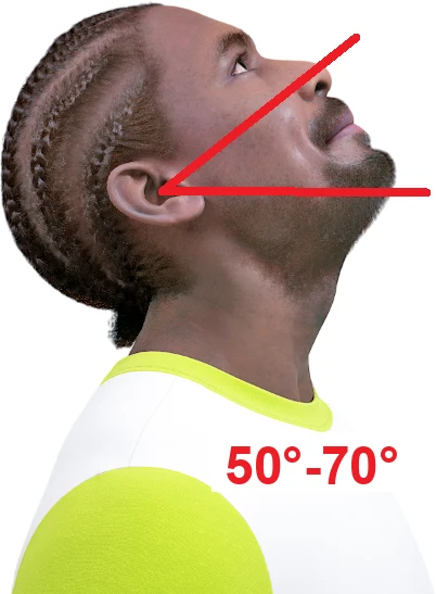
- Extension tilts the head backward, as in looking up at the ceiling.
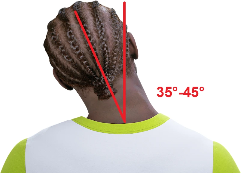
- Lateral flexion bends the head sideways, ear toward shoulder.
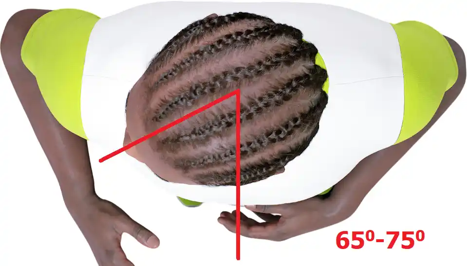
- Rotation turns the head side to side, as in looking over a shoulder.
What is a normal range of motion for the neck?
Clinical reference charts, the kind used in physical exams, typically list these ranges for an adult:
| Movement | Typical clinical range |
|---|---|
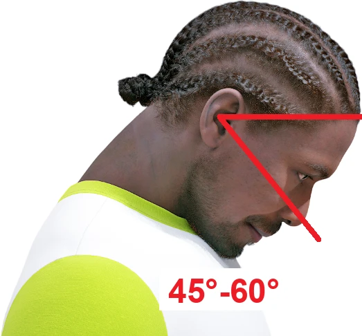
| Flexion (chin to chest) | 45°-60° |
| Extension (looking up) | 50°-70° |
| Rotation (looking over the shoulder), each side | 65°-75° |
| Lateral flexion (ear to shoulder), each side | 35°-45° |
These bands run wider than a single textbook number because published reference values disagree with each other, especially for extension, where some older clinical charts run noticeably higher than what measured studies in healthy adults actually find.1 A 2023 study that measured 300 healthy adults aged 18 to 29 with a digital goniometer found averages of 57.7 degrees of flexion, 59.1 degrees of extension, about 70 degrees of rotation to each side, and about 42 degrees of lateral flexion to each side, all inside the ranges above.2 A chart like this is a reference band, not a pass-or-fail line: what matters more day to day is whether your own mobility has changed, and whether it’s roughly symmetrical side to side.
Each of the four movements draws on the AO joint, the AA joint, and the lower cervical spine in different proportions.
Cervical flexion (chin to chest)
Flexion is the forward, downward movement of the head toward the chest. Most of it happens lower in the neck:
| Joint | Flexion contribution |
|---|---|
| AO (C0-C1) | 5°-10° |
| AA (C1-C2) | 5°-10° |
| Lower cervical spine (C3-C7) | 35°-40° |
Cervical extension (looking up)
Extension is the backward, upward movement of the head, as in looking up at the ceiling. Like flexion, the lower cervical spine carries most of it:
| Joint | Extension contribution |
|---|---|
| AO (C0-C1) | ≈10° |
| AA (C1-C2) | ≈10° |
| Lower cervical spine (C3-C7) | 30°-50° |
Cervical rotation (turning the head sideways)
Rotation turns the head to look over one shoulder without moving the torso. Here the split is reversed, the AA joint does most of the work:
| Joint | Rotation contribution |
|---|---|
| AO (C0-C1) | negligible |
| AA (C1-C2) | 35°-40° |
| Lower cervical spine (C3-C7) | 30°-35° |
That AA figure lines up with independent anatomy references, which put atlanto-axial rotation at roughly 40 degrees (a reported range of 39-49 degrees) and its share of total neck rotation at 50-60 percent, more than any other single segment.34 Because the AA joint carries so much of the load, a rotation deficit and a flexion or extension deficit often point to different regions of the neck, useful context when you’re trying to figure out where stiffness is actually coming from.
Cervical lateral flexion (tilting the head to the side)
Lateral flexion tilts the head toward the shoulder, as if trying to touch an ear to it. It’s the smallest of the four ranges, and mostly a lower-cervical movement, with the AO joint contributing only a few degrees on its own.5
| Joint | Lateral flexion contribution |
|---|---|
| AO (C0-C1) | 5°-8° |
| AA (C1-C2) | minimal (5°-10°) |
| Lower cervical spine (C3-C7) | 25°-30° |
A few patterns repeat across all four tables: the lower cervical spine contributes the most to flexion, extension, and lateral flexion; the AO joint plays almost no role in rotation; and the AA joint alone accounts for roughly half of the neck’s total rotation. Read together, they’re a reference point for whether a given restriction fits a normal pattern or looks unusual enough to have checked.
How is neck range of motion measured, in a clinic and at home?
In a clinic or physical therapy setting, cervical range of motion is usually read with a goniometer or an inclinometer, a hinged, protractor-style tool aligned with the head and shoulders to capture the angle at the end of a movement. Some settings use a device that straps to the head and tracks the angle digitally through the full motion, closer to how the 2023 goniometer study above captured its normative data.2 The instrument matters less than consistency: measuring the same way, at the same time of day, is what makes a follow-up reading meaningful.
You won’t get clinical precision without the right equipment, but a rough self-check is useful for tracking your own trend over time. Three simple options:
Mirror-based estimate
Stand facing a mirror, or mark a reference point on a wall at eye level. Move through each direction and compare the angle by eye against the tables above, using your ear, chin, or nose as the reference point.
Smartphone apps
Several free apps use a phone’s built-in gyroscope as a digital inclinometer: hold the phone flat against your forehead or the crown of your head, and the app reads the angle as you move through each direction. It’s not a substitute for professional testing, but it’s more consistent than eyeballing a mirror for tracking change over a few weeks.
Functional range test
No angle reading needed, just a quick check of whether each movement feels complete and roughly symmetrical: try to touch your chin to your chest, look up at the ceiling without discomfort, look over each shoulder, and bring each ear toward the matching shoulder. Note any side that feels noticeably more restricted than the other.
Why might you lose range of motion in your neck?
A gradual drop in range of motion is usually tied to daily habits and repetitive patterns rather than a single dramatic event. Identifying which one applies is the first step toward restoring mobility.
- Posture and a sedentary routine: hours spent looking down at a phone or hunched toward a monitor, so-called tech neck, keep the cervical spine parked in a flexed position. Reading for long stretches with the head down, and long periods of driving or sitting with limited movement, add to the same pattern.
- Sleep position and pillow choice: a pillow that’s too high, too flat, or otherwise misaligns the neck overnight can leave it stiff by morning, especially after sudden movements during sleep or sleeping with the head turned to one side for an extended stretch.
- Repetitive or asymmetrical activity: cycling holds the head in a fixed position for long periods; walking on uneven terrain asks the neck to make constant small corrections; sewing, sketching, or writing keeps the head down and repeats the same arm movement; carrying a bag on one shoulder unevenly loads the neck and upper back; and sports that favor one side of the body, tennis, golf, or baseball among them, can build muscle imbalances that restrict motion more on one side than the other.
- Age-related change: discs and joints lose some of their give over the decades, a normal part of how the spine changes rather than an injury in itself. It typically shows up as a slow, symmetrical narrowing of range rather than a sudden loss.
- Injury: a sudden impact, a fall, or a strain from lifting or moving awkwardly can cause a sharper, more one-sided drop in mobility, often paired with tenderness or swelling.
If your neck stiffness persists despite stretching and posture changes, or if a drop in range of motion comes with numbness, tingling, or weakness radiating into an arm or hand, that’s a reason to see a healthcare provider rather than to keep self-managing with stretching alone.
Where can you go from here?
We put the flexion, extension, rotation, and lateral flexion reference angles from this guide onto a single printable page: download the Neck Range of Motion Exercises PDF to keep next to your desk or gym bag as a quick visual check.
For the exercises themselves, chin tucks, stretches for each of the four movements, and how to progress them, see our companion guide, 7 best stiff neck stretches for pain relief. Once bodyweight stretching feels controlled and pain-free, a tool with adjustable resistance, such as the Bejoyland NECKY, lets you add traction and load to the same movements for static, dynamic, or PNF-style work, building the mobility gains from stretching into more durable strength.
NECKY and the resources above are built for general fitness, mobility, and posture support, not to diagnose or treat a medical condition. Individuals with pre-existing cervical-spine conditions should consult a healthcare provider or therapist before starting a training program.
FAQ
What is a normal range of motion for the neck?
Clinical charts generally put normal cervical range of motion at 45-60 degrees of flexion, 50-70 degrees of extension, 65-75 degrees of rotation to each side, and 35-45 degrees of lateral flexion to each side, with some variation depending on age, sex, and how it’s measured.
What is the correct ROM for the neck?
There is no single “correct” number, only a typical range, since normal values vary by age, sex, and measurement method. A 2023 goniometer study of 300 healthy adults found averages around 58 degrees of flexion, 59 degrees of extension, 70 degrees of rotation, and 42 degrees of lateral flexion, all within the broader clinical range.
What is the total range of motion of the human neck?
Adding up the four movements gives a rough picture: about 45-60 degrees of flexion plus 50-70 of extension for a combined 95-130 degrees of forward-to-back motion, and about 65-75 degrees of rotation to each side, roughly 130-150 degrees side to side, though clinicians rarely add the numbers this way since each movement is assessed on its own.
Why have I lost range of motion in my neck?
Most gradual loss traces back to posture and screen habits, sleep position and pillow choice, repetitive or one-sided movement, or age-related change in the joints and discs. A sudden, one-sided drop after an injury is different, so have it checked by a healthcare provider.
Which joint contributes the most to neck rotation?
The atlanto-axial (AA) joint between C1 and C2 supplies roughly 50-60 percent of the neck’s total rotation on its own, more than any other single segment of the cervical spine.
Can you measure your own neck range of motion?
Yes, with a mirror, a phone’s gyroscope-based inclinometer app, or a simple functional test like touching your chin to your chest and looking over each shoulder. It won’t match clinical precision, but it’s enough to track whether your own mobility is improving or declining.
Sources
- Swartz EE, Floyd RT, Cendoma M. Cervical Spine Functional Anatomy and the Biomechanics of Injury Due to Compressive Loading. Journal of Athletic Training, 2005. PMC.
- Apti M, Kuru Colak T, Akcay B. Normative Values for Cervical and Lumbar Range of Motion in Healthy Young Adults. The Journal of Turkish Spinal Surgery, 2023.
- Kenhub. Atlantoaxial joint: Anatomy, function, movements. Kenhub Anatomy Library.
- Gonzalez DCN, et al. C1-C2 Rotatory Subluxation in Adults: A Narrative Review. Diagnostics, 2022. PMC.
- Kenhub. Atlanto-occipital joint: Bones, ligaments, movements. Kenhub Anatomy Library.